In my recent posts, we showed different experiments with C19 vaccinated blood.
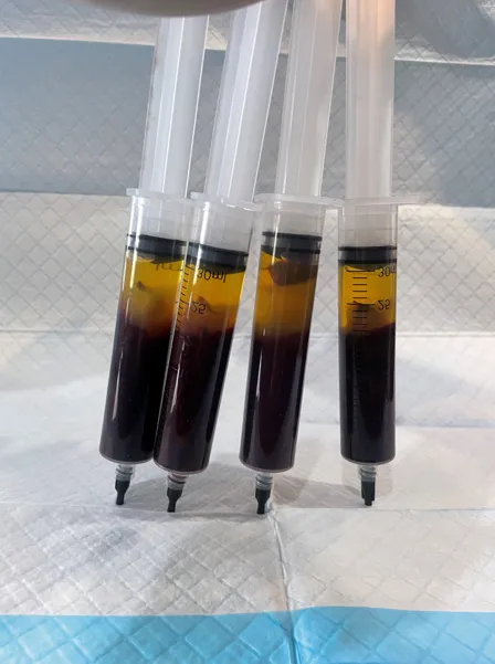
Image: Four vaccinated Blood Samples left to sit for 4 hours I have posted the work of Clifford Carnicom and myself showing that Near Infrared Spectroscopy performed on C19 vaccinated and unvaccinated blood repetitively identified the spectral signature of functional chemical groups indicating polymer hydrogels.
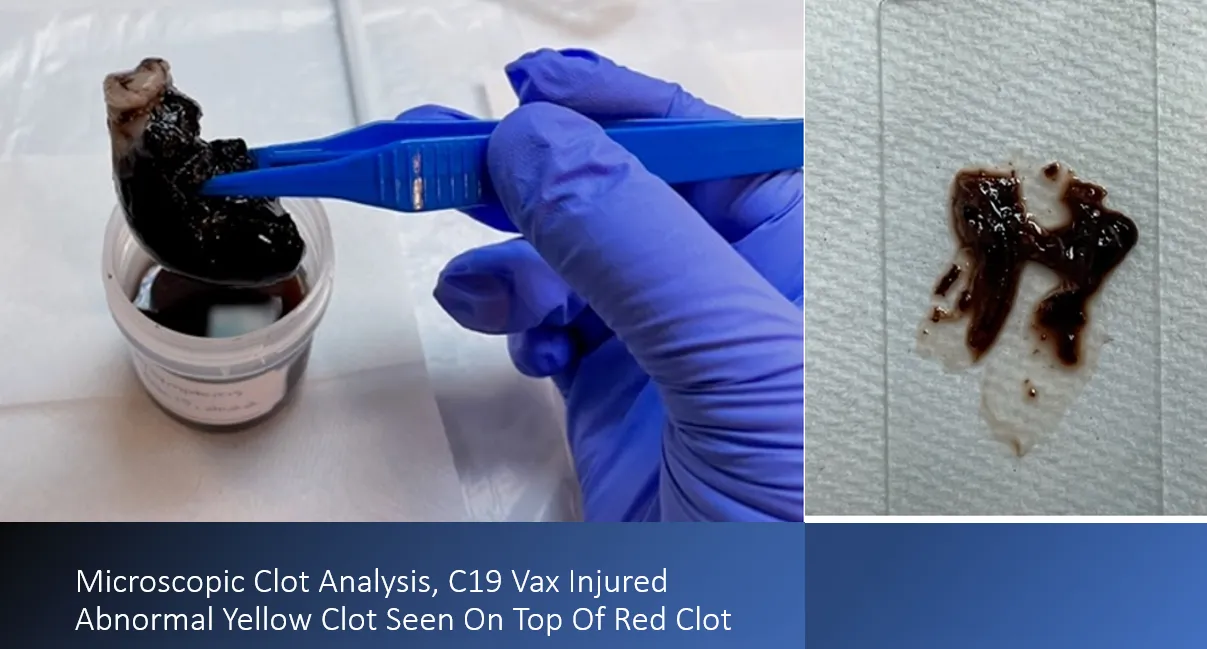
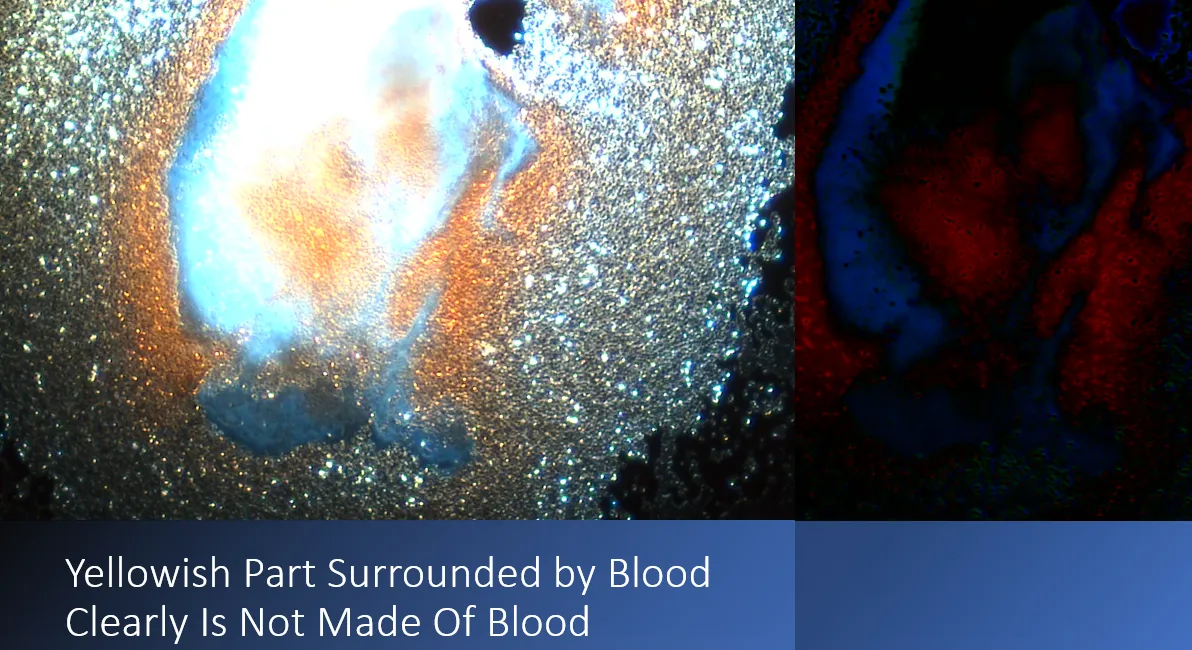
In this article, I continue the investigation and analyze one of the blood clots from a C19 vaccine injured individual. The blood clot was received in Formalin. In the image above, the clot was removed from the container. Two slides were prepared, one analyzing tissue from the red part of the clot, the other from the upper yellow part of the clot. This is what I found:
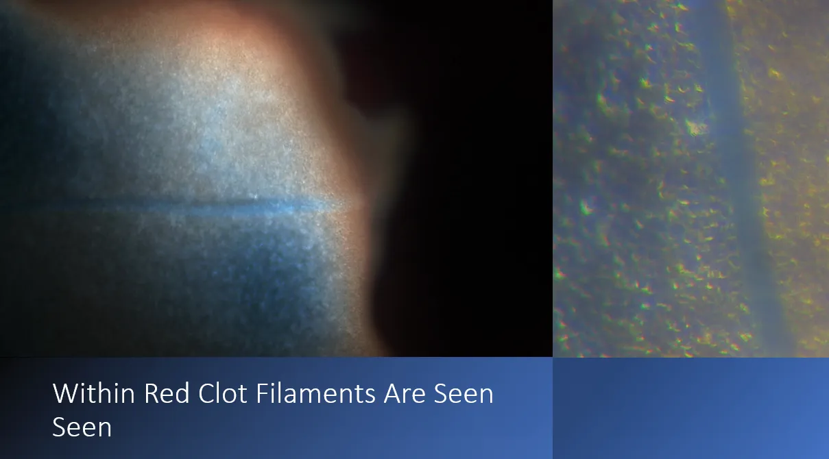
In this video I show how long the blue filament is running within the red part of the clot. This is 100x magnification.


The blue and white filaments have been seen in the C19 Vials and in Live blood.
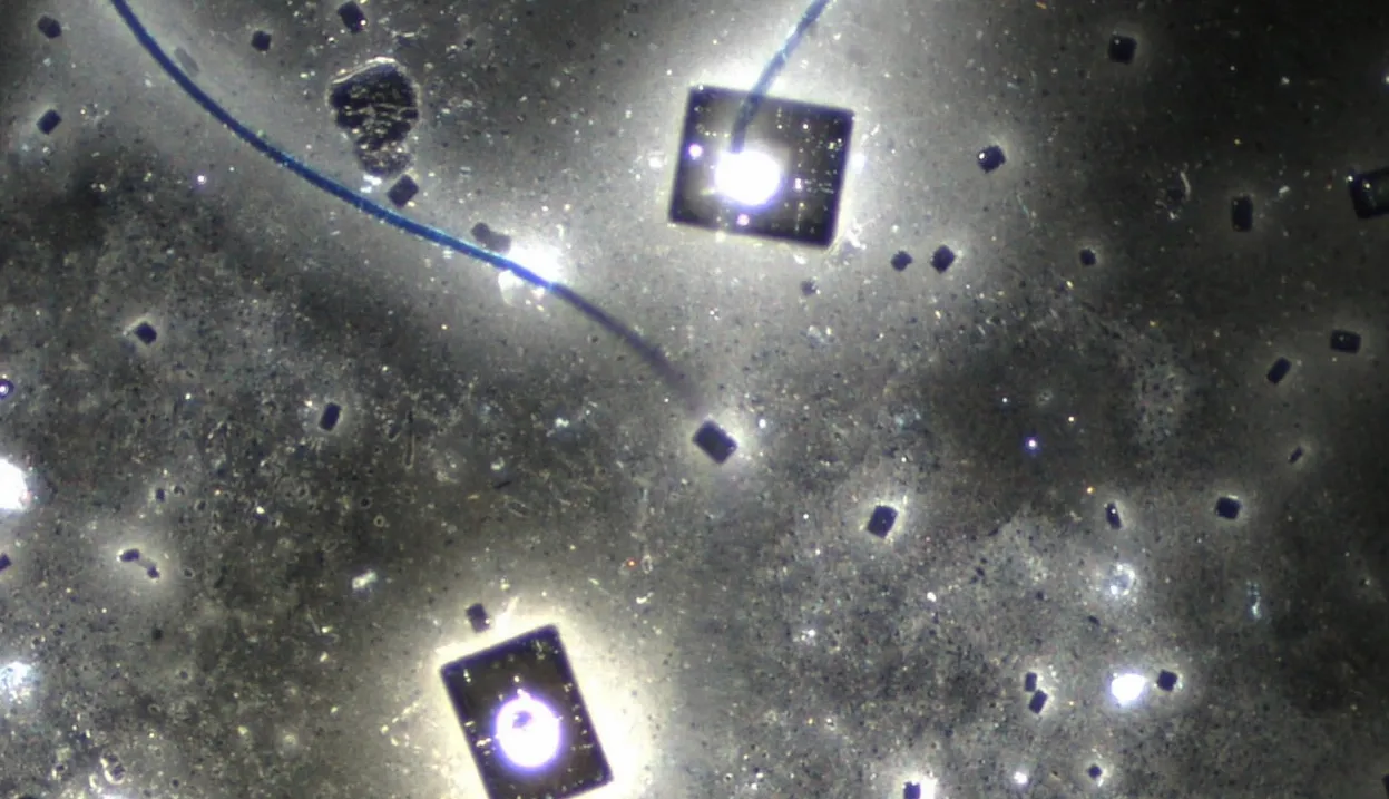
Image: Dr. David Nixon. C19 Pfizer Vial Contents. Darkfield Microscopy, 200x
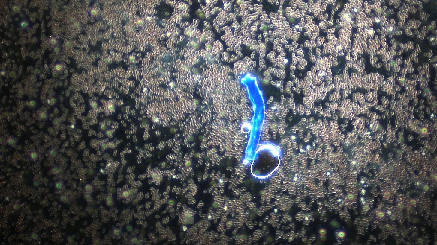
Image: Unvaccinated Blood, Dr. Ana’s office. Blue Filament. 100x magnification
Summary:
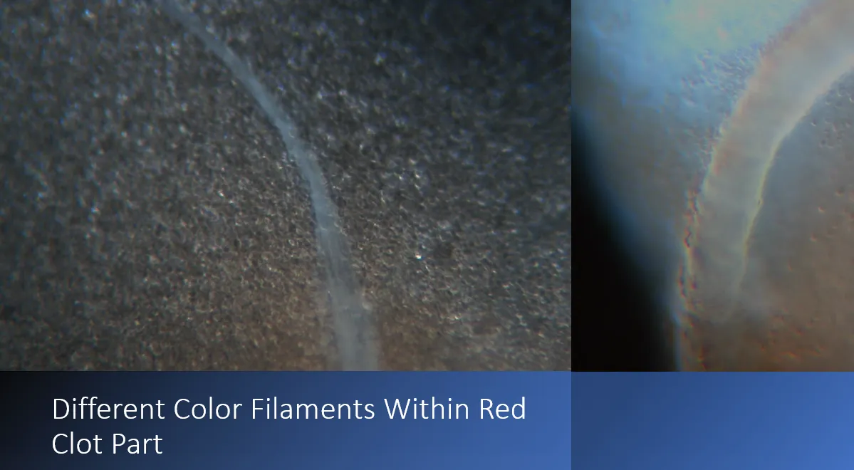
The red blood clot part had classic filaments that we see in live blood now in vaccinated and unvaccinated individuals. The upper rubbery part of the clot looks like sheets of a material – it is not made of blood cells. I had showed the video in my above linked post that showed this clot to be very rubbery, and not dissolvable by mechanical means. Further analysis is needed and will follow.
Tags:
01. Unwanted Medical Procedures,
05. Informed Consent,
06. Product and Treatment Safety,
09. Patient Centric Care,
12. Civil Liberties Protections,
14. Limits to Government Powers,
16. Protected Medical Class,
18. Criminal Penalties,
22. Employee Protection,
23. Consumer Protections,
24. Healthcare Provider Education,
Articles,
Blood Clots,
Downstream Problems,
Employer Mandated Vaccinations,
Important,
Informed Consent,
MFA Articles,
Mandates,
Mass Vaccinations,
Medical Tyranny,
Moderna,
Pfizer BioNTech,
Pfizer Comirnaty,
Plandemic,
Problems,
Restrictions,
Statistics,
Vaccine Deaths,
Vaccine Hesitancy,
Vaccine Injury,
mRNA Vaccine